Condyloma acuminata (CA), also known as anogenital warts, are benign proliferative epidermal and/or mucosal lesions usually caused by Human Papilloma Virus (HPV) type 6 and 11. They initially manifest as variable sized and shaped soft papules or plaques on anogenital skin. However, they can grow as a large, bulky, lobulated growth. Lesions are commonly multiple and multifocal, affecting the vulva, perianal, vaginal and cervical regions. They represent the most common sexually transmitted disease (STD) and are highly contagious. Further, the incidence of CA is 5-7fold higher in Human Immunodeficiency virus (HIV) positive patients compared to immunocompetent patients. The HIV infection reduces the local immune control of HPV infection thus favours the proliferation of the HPV which results into large sized CA. The giant condyloma acuminata (GCA) and giant condyloma of Buschke-Löwenstein (GCBL) are uncommon variants of classical condyloma acuminata (CCA) which can reach the size of 10 to 30 cm. The treatment of CA should be individualized and based upon the extent of disease and treatment availability. The small CA which present as papules or plaques can be treated by Podophyllin, Imiquimod, electrosurgical ablation or cryotherapy. However, GCA require excision with cold knife, electrosurgery or CO2 laser. When the base of GCA is narrow, surgical excision with minimal skin loss allows primary closure of the wound, whereas when the base is broad and relatively fixed, one must keep the differential diagnoses of GCA versus GCBL which require wide excision and reconstruction of the tissue. This case report illustrates the surgical management of GCA with broad base in a 44-year-old female patient with HIV infection. She was treated by wide surgical excision followed by reconstruction of the defect with fascio-cutaneous V-Y advancement flaps. Histology confirmed the diagnosis of condyloma acuminatum. Six months following surgery, she had recurrence of a perianal wart that was cauterized. On further 12 month follow up there was no new recurrence of any condylomatous lesion.
This is an Open Access article, distributed under the terms of the Creative Commons Attribution 4.0 International License (http://creativecommons.org/licenses/by/4.0/), which permits unrestricted use, distribution and reproduction in any medium or format, provided the original work is properly cited.
Human Papilloma Virus, Giant Condyloma Acuminata, Giant Condyloma of Buschke-Löwenstein, Bushcke-Lowenstein Tumour
1. Introduction
Condyloma acuminata (CA) also known as anogenital or venereal warts is a common sexually transmitted disease (STD) among females and males of reproductive age group
[1]
Athanase L, Bonaventura CTM, Abdallah M, Dismas M, Albert K, Balthazar G. Giant condyloma ccuminatum of vulva in an HIV-infected woman. Case Reports in Infectious Diseases. Volume 2017, Article ID 5161783, 3pages.
.The causal role of Human papillomavirus (HPV) is well established in CA
[1]
Athanase L, Bonaventura CTM, Abdallah M, Dismas M, Albert K, Balthazar G. Giant condyloma ccuminatum of vulva in an HIV-infected woman. Case Reports in Infectious Diseases. Volume 2017, Article ID 5161783, 3pages.
Adebajo SB, Nowak RG, Adebiyi R, Shoyemi E, Ekeh C, Ramadhani HO, et al. Prevalence and factors associated with anogenital warts among sexual and gender minorities attending a trusted community health center in Lagos, Nigeria. PLOS Glob Public Health. 2022; 2(11): e0001215.
. More than 90% of cases of CA are caused by low-risk, non-oncogenic HPV type 6 and/or 11
[9]
Adebajo SB, Nowak RG, Adebiyi R, Shoyemi E, Ekeh C, Ramadhani HO, et al. Prevalence and factors associated with anogenital warts among sexual and gender minorities attending a trusted community health center in Lagos, Nigeria. PLOS Glob Public Health. 2022; 2(11): e0001215.
Low AJ, Clayton T, Konate I, Nagot N, Ouedraogo A, Huet C et al. Genital warts and infection with human immunodeficiency virus in high-risk women in Burkina Faso: a longitudinal study. BMC Infectious Diseases 2011; 11(20): 1471-2334.
[11]
Dareng EO, Adebamowo SN, Famooto A, Olawande O, Odutola MK, Olaniyan Y et al. Prevalence and incidence of genital warts and cervical Human Papillomavirus infections in Nigerian women. BMC Infectious Diseases. 2019; 19(27)
The worldwide prevalence of CA is estimated at 0.13%-5.1% with an incidence of 76-190 per 100,000 person-years among women
[9]
Adebajo SB, Nowak RG, Adebiyi R, Shoyemi E, Ekeh C, Ramadhani HO, et al. Prevalence and factors associated with anogenital warts among sexual and gender minorities attending a trusted community health center in Lagos, Nigeria. PLOS Glob Public Health. 2022; 2(11): e0001215.
. However, the incidence is 5-7 times higher in Human Immunodeficiency virus (HIV) positive patients
[10]
Low AJ, Clayton T, Konate I, Nagot N, Ouedraogo A, Huet C et al. Genital warts and infection with human immunodeficiency virus in high-risk women in Burkina Faso: a longitudinal study. BMC Infectious Diseases 2011; 11(20): 1471-2334.
[11]
Dareng EO, Adebamowo SN, Famooto A, Olawande O, Odutola MK, Olaniyan Y et al. Prevalence and incidence of genital warts and cervical Human Papillomavirus infections in Nigerian women. BMC Infectious Diseases. 2019; 19(27)
.There is a complex interaction between HIV and HPV, and local mucosa’s immunological mechanisms. HIV increases HPV transcription, and it causes a decrease in the number of macrophages, Langerhans cells and CD4 T lymphocytes in the mucosa with a consequent reduction in local immune control of HPV infection and increased proliferation of the virus. The HPV viral DNA is incorporated into cellular DNA redirecting cellular processes into dysregulated cellular proliferation
[9]
Adebajo SB, Nowak RG, Adebiyi R, Shoyemi E, Ekeh C, Ramadhani HO, et al. Prevalence and factors associated with anogenital warts among sexual and gender minorities attending a trusted community health center in Lagos, Nigeria. PLOS Glob Public Health. 2022; 2(11): e0001215.
The giant condyloma acuminata (GCA) and giant condyloma of Buschke-Löwenstein (GCBL) also known as Buschke-Löwenstein tumor (BLT) are uncommon variants of classical condyloma acuminatum (CCA). They are slow growing tumour and can grow to 10-30cm in size. Although histopathologically benign, they have a potential to grow into squamous cell carcinoma (SCC), and have a high recurrence rate
[14]
Nieves-Condoy JF, Acuña-Pinzón CL, Chavarría-Chavira JL, Hinojosa-Ugarte D, Zúñiga-Vázquez LA. Giant condyloma acuminata (Buschke-Lowenstein Tumor): Review of an unusual disease and difficult to manage. Infectious Diseases in Obstetrics and Gynecology. Volume 2021, Article ID 9919446, 5 pages
Tas S, Arik MK, Ozkul F, Cikman O, Akgun Y. Perianal giant condyloma acuminatum—Buschke-L¨owenstein tumor: A case report. Case Reports in Surgery. Volume 2012, Article ID 507374, 3 pages
. Most authors recommend wide surgical excision as the treatment of choice which allows a complete histological examination and assessment of tumor-free resection margins
[1]
Athanase L, Bonaventura CTM, Abdallah M, Dismas M, Albert K, Balthazar G. Giant condyloma ccuminatum of vulva in an HIV-infected woman. Case Reports in Infectious Diseases. Volume 2017, Article ID 5161783, 3pages.
Peri Eriad Y, Chaidir AM, Agus Rizal AHH, Chaula LS, Rainy U. Surgical management of giant genital condyloma acuminata by using double keystone flaps. Case Reports in Urology. Volume 2016, Article ID 4347821, 5pages.
Tas S, Arik MK, Ozkul F, Cikman O, Akgun Y. Perianal giant condyloma acuminatum—Buschke-L¨owenstein tumor: A case report. Case Reports in Surgery. Volume 2012, Article ID 507374, 3 pages
A 44-year-old, para 2 presented to gynaecology clinic with a history of vulval growth for three years. It started as a small warty growth which continued to grow in size. It was associated with itching, malodourous discharge and sometimes mild bleeding specially on rubbing. She had difficulty in walking and sitting.
She attained early menopause at the age of 42 years and was experiencing mild hot flushes. She was diagnosed with HIV infection six years ago and was then started on highly active anti-retroviral treatment (HAART). However, she was switched to second line HAART regimen two years ago due to virological failure which included Aluvia, Abacavir and Lamivudine. She was not vaccinated for HPV. The Pap smear showed low grade squamous intraepithelial lesion. She did not have any other comorbidity.
On examination, she was of average build. Vital signs were normal with a pulse of 70 beats per minute and BP of 130/60 mm of Hg. Her temperature was normal.
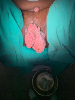
There were huge multiple exophytic masses, cauliflower-like in appearance, pink and grayish in colour involving both labia majora and minora. They extended anteriorly to mons pubis and posteriorly to the perineum and perianal area. They measured 15 x 7 x 5 cm on the left and 11x 6 x 5cm the on right side. The masses were soft to firm in consistency, friable with irregular surface and had a broad base (4-5cm) but were not fixed to underlying structures. The margins were well defined. Some areas of the masses were infected and were draining foul smelling yellow discharge. There were a few satellite lesions on the mons pubis and on the right groin. Bilateral inguinal lymph nodes were enlarged to 0.5 cm, discrete and mobile and seemed to be reactive lymph nodes.
The laboratory investigations showed haemoglobin of 10gm/dl, platelet count of 267 x 109/L, and white cell count of 9.2 x 10⁹/L. The CD4 count was 285 cell/mm3 and viral load was 35 copies/ml. The urea was 3.7mmol/L and creatinine was 8.3umol/L with normal electrolyte panel. The liver function tests were within normal range except mild hypoalbuminemia of 31 gm%. A clinical diagnosis of GCA with HIV on second line of HAART was made. Multiple biopsies from different sites were taken in the ward which confirmed the diagnosis of CA.
Figure 2. Pre-operative image of GCA from another angle.
Patient was advised to take Sitz bath and was empirically given oral Sulfamethoxazole + Trimethoprim to control the local infection. She was started on Imiquimod cream 5% three times a week for three months while awaiting surgery. The infection had cleared at the time of surgery but there was no appreciable regression in the size of the growth.
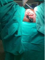
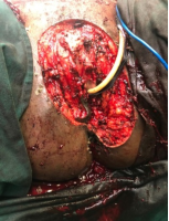
Given the broad base of the condyloma, it was anticipated that there would be wide skin loss; hence, the management was planned with the plastic surgeon. The patient underwent simple vulvectomy. The skin with superficial subcutaneous tissue of the part of the mons pubis, clitoris, both labia majora and minora, left side of perineum and perianal area were resected en-block with a margin of 1.0 cm and sent for histology, followed by reconstruction of the vulva with a fascio-cutaneous V-Y advancement flaps from both thighs. One satellite lesion on the mons pubis was excised and others were cauterized.
Histology showed hyperkeratosis, parakeratosis, acanthosis, excessive koilocytes in the epidermis and congested vessels in the dermis which were in keeping with the features of condyloma acuminatum and chronic inflammation with no evidence of high-grade dysplasia or invasive malignancy. The resection margins were clear of disease. The histological diagnosis was in keeping with GCA rather than GCBL as argued in the discussion section of the report.
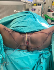
The post-operative course was complicated by infection of the wound which was successfully treated with intravenous Tazocin x 4.5 gm x eight hourly for ten days and wound care. The patient was discharged on day 21. Both vulval and thigh wounds had healed well and flaps were well taken up. Six months later the patient had recurrence of a perianal wart which was cauterized. Since then, she remained free of recurrence at 12 months follow-up and had no complaints.
Figure 5. Six months following vulvectomy and vulval reconstruction.
3. Discussion
HPV is one of the most common STD, with a lifetime risk of 50-80%
[6]
Thaker BD, Bhardwaj S. Giant condyloma acuminatum of vulva-a case report. 2017: 6(2) • Issn No 2277 - 8179 | If: 3.508 | Ic Value: 78.46.
[6]
. There are more than 200 types of HPV, some are oncogenic others are non-oncogenic strains
[11]
Dareng EO, Adebamowo SN, Famooto A, Olawande O, Odutola MK, Olaniyan Y et al. Prevalence and incidence of genital warts and cervical Human Papillomavirus infections in Nigerian women. BMC Infectious Diseases. 2019; 19(27)
.The highest risk of contracting HPV infection is between 18-28 years
[6]
Thaker BD, Bhardwaj S. Giant condyloma acuminatum of vulva-a case report. 2017: 6(2) • Issn No 2277 - 8179 | If: 3.508 | Ic Value: 78.46.
[6]
.Although, 90% of infected people are able to clear HPV by strong localized cell mediated immune response, approximately 10% of individuals develop persistent infection. The persistent infection may result in benign proliferative lesions such as CCA, GCA, GCBL/BLT. The majority of the cases of benign lesions are caused by non-oncogenic strains mainly 6 and 11
Peri Eriad Y, Chaidir AM, Agus Rizal AHH, Chaula LS, Rainy U. Surgical management of giant genital condyloma acuminata by using double keystone flaps. Case Reports in Urology. Volume 2016, Article ID 4347821, 5pages.
Meites E, Gee J, Elizabeth Unger E, Markowitz L. Human papilloma virus. 4thedition. Epidemiology and Prevention of vaccine-Preventable Diseases. Public Health Foundation. 168-1781. Downloaded from URL:
In the populations where HIV prevalence is high in adults of reproductive age such as in South Africa (about 19.5%), there appears to be higher incidence of GCA
[5]
Moodley M, Govender PS. External beam radiotherapy for large genital warts: Does it work?. Eur. J. Gynaecol. Oncol. - ISSN: 0392-2936 XL, n. 2, 2019
GCA and GCBL are uncommon variants of CCA. In 1925, Buschke and Lowenstein described a unique neoplasm of the penis which, to them, bore similarities to CCA as well as SCC. Clinically it looked like CA but behaved like locally invasive malignancy, whereas histologically it was more like condyloma acuminatum without any evidence of anaplasia
[12]
Boda D, Cutoiu A, Bratu D, Bejinariu N, Crutescu R. Buschke-Löwenstein tumors: A series of 7 case reports. Experimental and Therapeutic Medicine. 2022; 23: 393-403
Given the rarity of the disease, the literature is available as individual case reports or series of case reports. And in the available literature, most of the clinical reports have not made any differentiation between GCA and GCBL/BLT rather have used the terms interchangeably, but a few, especially the pathology reports have made a distinction between the two.
Although, the GCA and GCBL/BLT are often used mutually, it is prudent to compare and contrast the two as it has clinical implications. Both GCA and GCBL have similar risk factors including immunosuppression, diabetes, smoking, multiple partners, men having sex with men (MSM), poor hygiene as well as other STDs
[7]
Mittal P, Prakash V, Gupta R, Dewan R, Singhal S, Suri J. Giant condyloma acuminatum of vulva treated by surgical excision and reconstruction of defect. Arch Gynecol Obstet 2013; 287: 1047-48.
[12]
Boda D, Cutoiu A, Bratu D, Bejinariu N, Crutescu R. Buschke-Löwenstein tumors: A series of 7 case reports. Experimental and Therapeutic Medicine. 2022; 23: 393-403
. They have comparable clinical presentation and management but the risk of malignant transformation, recurrence rates and prognosis are different. The current patient had HIV disease which is a risk factor for both GCA and GCBL.
The risk of transformation into SCC in GCBL ranges from 30% to 56%, the risk is assumed to be less in GCA, whereas CCA has a small risk of 2%. The risk increases when there is mixed infection with oncogenic strains of HPV
[5]
Moodley M, Govender PS. External beam radiotherapy for large genital warts: Does it work?. Eur. J. Gynaecol. Oncol. - ISSN: 0392-2936 XL, n. 2, 2019
Tas S, Arik MK, Ozkul F, Cikman O, Akgun Y. Perianal giant condyloma acuminatum—Buschke-L¨owenstein tumor: A case report. Case Reports in Surgery. Volume 2012, Article ID 507374, 3 pages
The GCBL recurrence rate can be as high as 60-66% which largely depends on the spread of the tumour and (in)adequacy of the resection. Whereas the recurrence rate in adequately resected GCA is 13-29%
[14]
Nieves-Condoy JF, Acuña-Pinzón CL, Chavarría-Chavira JL, Hinojosa-Ugarte D, Zúñiga-Vázquez LA. Giant condyloma acuminata (Buschke-Lowenstein Tumor): Review of an unusual disease and difficult to manage. Infectious Diseases in Obstetrics and Gynecology. Volume 2021, Article ID 9919446, 5 pages
Tas S, Arik MK, Ozkul F, Cikman O, Akgun Y. Perianal giant condyloma acuminatum—Buschke-L¨owenstein tumor: A case report. Case Reports in Surgery. Volume 2012, Article ID 507374, 3 pages
Both GCA and GCBL look alike; bulky, cauliflower-shaped masses that can grow from 5cm to 20 to 30cm that cause physical symptoms, emotional distress and adversely affect quality of life but there are subtle differences in the clinical behaviour and histological features of the two.
GCA and GCBL both are largely located on the genital area (vulva in females and penis in males) and may extend to the perineum and perianal area. The anorectal region is the second common site for GCBL with a propensity for perianal fistula formation and infection. GCBL has broad base and fixed to underlying structures to variable degree, whereas GCA can have narrow or broad base but mostly not fixed to underlying structures
Histologically, GCA and GCBL show many similar characteristic features such as orderly arrangement of the epithelial layers, well-formed papillae with a prominent central fibrovascular core, hyperkeratosis with parakeratosis and marked acanthosis, koilocytes and chronic inflammatory infiltration. There is no lymphatic, vascular or neuronal invasion, and the basement membrane integrity is maintained. Both do not metastasize, neither is there atypia of cells
[4]
Peri Eriad Y, Chaidir AM, Agus Rizal AHH, Chaula LS, Rainy U. Surgical management of giant genital condyloma acuminata by using double keystone flaps. Case Reports in Urology. Volume 2016, Article ID 4347821, 5pages.
. However, the major differentiating point between the two is that the GCA is largely an exophytic growth whereas GCBL has both exophytic as well as endophytic growth. Typically, GCBL causes pushing infiltration of underlying dermis and deeper structure with an intact basement membrane. The invasive strands of the tumour have a well-developed basal cell layer of epidermis
[13]
Manzione TD, Nadal SR, Veasey J V. Perianal Buschke-Lowenstein tumor: report of two cases treated with 25% podophyllin ointment. Surg Cosmet Dermatol. Rio de Janeiro. 2020; 12(S1): 58-61.
[14]
Nieves-Condoy JF, Acuña-Pinzón CL, Chavarría-Chavira JL, Hinojosa-Ugarte D, Zúñiga-Vázquez LA. Giant condyloma acuminata (Buschke-Lowenstein Tumor): Review of an unusual disease and difficult to manage. Infectious Diseases in Obstetrics and Gynecology. Volume 2021, Article ID 9919446, 5 pages
. Nevertheless, the endophytic growth causes invasion and destruction of the underlying and adjacent tissues simulating the malignant invasion
[12]
Boda D, Cutoiu A, Bratu D, Bejinariu N, Crutescu R. Buschke-Löwenstein tumors: A series of 7 case reports. Experimental and Therapeutic Medicine. 2022; 23: 393-403
There is a changing concept over time that not alllarge (>3cm) and giant (>10cm) condylomas are GCBL, and it is considered that the CCA, GCA, GCBL are the continuum of the same benign entity but not mandatory spectrum of potentially malignant lesions
[12]
Boda D, Cutoiu A, Bratu D, Bejinariu N, Crutescu R. Buschke-Löwenstein tumors: A series of 7 case reports. Experimental and Therapeutic Medicine. 2022; 23: 393-403
The patient described in this report fits into the criteria of GCA, not GCBL as it was giant in size (>10cm), and it had exophytic growth only, and both clinically and histologically there was no evidence of extrusion of tumour in the underlying structures.
The treatment options for CCA are topical therapy (podophyllin, 5-FU, Imiquimod, intra-lesional injections of interferons, ablative therapy (cryotherapy, electrosurgery) and excisional therapy with CO2 laser or cold knife
Karnes JB, Usatine RP. Management of external genital warts. Am Fam Physician. 2014; 90(5): 312-18.
[15]
Tas S, Arik MK, Ozkul F, Cikman O, Akgun Y. Perianal giant condyloma acuminatum—Buschke-L¨owenstein tumor: A case report. Case Reports in Surgery. Volume 2012, Article ID 507374, 3 pages
In case of GCA and GCBL, before treatment it is mandatory to do multiple site deep biopsies that will assist in making the diagnosis, confirmation or exclusion of the presence of dysplasia, microinvasion or invasive carcinoma and in treatment planning
[9]
Adebajo SB, Nowak RG, Adebiyi R, Shoyemi E, Ekeh C, Ramadhani HO, et al. Prevalence and factors associated with anogenital warts among sexual and gender minorities attending a trusted community health center in Lagos, Nigeria. PLOS Glob Public Health. 2022; 2(11): e0001215.
Herweijer E, Ploner A, Sparén P. Substantially reduced incidence of genital warts in women and men six years after HPV vaccine availability in Sweden. Vaccine. 2018; 36: 1917–20.
[9, 18]
.An MRI is warranted in cases where symptoms and signs suggest possible extrusion in the underlying or adjacent structures
[12]
Boda D, Cutoiu A, Bratu D, Bejinariu N, Crutescu R. Buschke-Löwenstein tumors: A series of 7 case reports. Experimental and Therapeutic Medicine. 2022; 23: 393-403
Tas S, Arik MK, Ozkul F, Cikman O, Akgun Y. Perianal giant condyloma acuminatum—Buschke-L¨owenstein tumor: A case report. Case Reports in Surgery. Volume 2012, Article ID 507374, 3 pages
A wide surgical excision with histologically clear margins is the mainstay of treatment of GCA and GCBL. The wide surgical excision poses a challenge for the reconstruction of tissue. Most case reports describe the skin thickness skin graft or flap to cover the surgical defect
[15]
Tas S, Arik MK, Ozkul F, Cikman O, Akgun Y. Perianal giant condyloma acuminatum—Buschke-L¨owenstein tumor: A case report. Case Reports in Surgery. Volume 2012, Article ID 507374, 3 pages
Herweijer E, Ploner A, Sparén P. Substantially reduced incidence of genital warts in women and men six years after HPV vaccine availability in Sweden. Vaccine. 2018; 36: 1917–20.
[15, 17, 18]
. In the index case, we did bilateral fascio-cutaneous V–Y advancement flaps to reconstruct the vulva.
Radiation therapy or chemo-radiation despite its neoadjuvant application in a limited number of GCA and GCBL, has largely been avoided, mostly because of the fear of causing further anaplastic transformations. Moreover, in a series of case reports of GCA the radiotherapy showed partial response in most cases, only a few had shown complete regression [5]. Radiotherapy should be reserved for patients who are not fit for surgery or when clear surgical margins are not attainable or, GCA or GCBL has foci of SCC
[9]
Adebajo SB, Nowak RG, Adebiyi R, Shoyemi E, Ekeh C, Ramadhani HO, et al. Prevalence and factors associated with anogenital warts among sexual and gender minorities attending a trusted community health center in Lagos, Nigeria. PLOS Glob Public Health. 2022; 2(11): e0001215.
Boda D, Cutoiu A, Bratu D, Bejinariu N, Crutescu R. Buschke-Löwenstein tumors: A series of 7 case reports. Experimental and Therapeutic Medicine. 2022; 23: 393-403
Imiquimod 5% cream seems to be ineffective; however, in some case reports, GCA has demonstrated a good response to Podophyllin which has an exfoliating, immunological, and antimitotic action. Weekly treatment with Podophyllin 25% in solid petroleum jelly for 4-6 months can result in complete regression of GCA
[13]
Manzione TD, Nadal SR, Veasey J V. Perianal Buschke-Lowenstein tumor: report of two cases treated with 25% podophyllin ointment. Surg Cosmet Dermatol. Rio de Janeiro. 2020; 12(S1): 58-61.
[13]
. Topical treatment with Podophyllin may be considered before surgery to reduce the size of GCA, thus facilitating resection and avoiding postoperative complications
[13]
Manzione TD, Nadal SR, Veasey J V. Perianal Buschke-Lowenstein tumor: report of two cases treated with 25% podophyllin ointment. Surg Cosmet Dermatol. Rio de Janeiro. 2020; 12(S1): 58-61.
[13]
.
4. Conclusion
Despite being an uncommon condition, the incidence of GCA is relatively higher in immunosuppressed patients. GCA must be differentiated from GCBL because GCBL has higher recurrence rates and higher malignant transformation index and higher mortality rates than GCA
[14]
Nieves-Condoy JF, Acuña-Pinzón CL, Chavarría-Chavira JL, Hinojosa-Ugarte D, Zúñiga-Vázquez LA. Giant condyloma acuminata (Buschke-Lowenstein Tumor): Review of an unusual disease and difficult to manage. Infectious Diseases in Obstetrics and Gynecology. Volume 2021, Article ID 9919446, 5 pages
. The wide local excision of the lesion with clear margins and reconstruction of the resultant skin defect gives good cosmetic and functional results in both GCA and GCBL
[1]
Athanase L, Bonaventura CTM, Abdallah M, Dismas M, Albert K, Balthazar G. Giant condyloma ccuminatum of vulva in an HIV-infected woman. Case Reports in Infectious Diseases. Volume 2017, Article ID 5161783, 3pages.
Mittal P, Prakash V, Gupta R, Dewan R, Singhal S, Suri J. Giant condyloma acuminatum of vulva treated by surgical excision and reconstruction of defect. Arch Gynecol Obstet 2013; 287: 1047-48.
[1, 2, 7]
.
Furthermore, in order to reduce the incidence of genital warts, the governments must invest in HPV vaccination programs. The randomized clinical trials and population-based studies have demonstrated that quadri-valent and nona-valent HPV vaccination of adolescents before sexual debut significantly decrease the incidence of genital warts
[11]
Dareng EO, Adebamowo SN, Famooto A, Olawande O, Odutola MK, Olaniyan Y et al. Prevalence and incidence of genital warts and cervical Human Papillomavirus infections in Nigerian women. BMC Infectious Diseases. 2019; 19(27)
Herweijer E, Ploner A, Sparén P. Substantially reduced incidence of genital warts in women and men six years after HPV vaccine availability in Sweden. Vaccine. 2018; 36: 1917–20.
[11, 18]
.
Abbreviations
BLT: Buschke-LöwensteinTumor
CA: Condyloma Accuminata
CCA: Classical Condyloma Accuminata
GCA: Giant Condyloma Acuminata
GCBL: Giant Condyloma of Buschke-Löwenstein
HAART: Highly Active Anti-Retroviral Treatment
HIV: Human Immunodeficiency Virus
HPV: Human Papilloma Virus
MSM: Men Having Sex with Men
SCC: Squamous Cell Carcinoma
STD: Sexually Transmitted Disease
Author Contributions
Asha Misra: Conceptualization, Writing - original draft
Puritan Madzhia: Writing - original draft
Thabo Malebana: Writing - review & editing
Dakalo Muavha: Writing - review & editing
John Boshomane: Writing - review & editing
Kaiser Baloyi: Writing - review & editing
Consent
The patient gave written informed consent for her case to be published with guarantee of confidentiality. All information as well as figures have been de-identified.
Conflicts of Interest
The authors declare no conflicts of interest.
References
[1]
Athanase L, Bonaventura CTM, Abdallah M, Dismas M, Albert K, Balthazar G. Giant condyloma ccuminatum of vulva in an HIV-infected woman. Case Reports in Infectious Diseases. Volume 2017, Article ID 5161783, 3pages.
Karnes JB, Usatine RP. Management of external genital warts. Am Fam Physician. 2014; 90(5): 312-18.
[4]
Peri Eriad Y, Chaidir AM, Agus Rizal AHH, Chaula LS, Rainy U. Surgical management of giant genital condyloma acuminata by using double keystone flaps. Case Reports in Urology. Volume 2016, Article ID 4347821, 5pages.
Thaker BD, Bhardwaj S. Giant condyloma acuminatum of vulva-a case report. 2017: 6(2) • Issn No 2277 - 8179 | If: 3.508 | Ic Value: 78.46.
[7]
Mittal P, Prakash V, Gupta R, Dewan R, Singhal S, Suri J. Giant condyloma acuminatum of vulva treated by surgical excision and reconstruction of defect. Arch Gynecol Obstet 2013; 287: 1047-48.
[8]
Meites E, Gee J, Elizabeth Unger E, Markowitz L. Human papilloma virus. 4thedition. Epidemiology and Prevention of vaccine-Preventable Diseases. Public Health Foundation. 168-1781. Downloaded from URL:
Adebajo SB, Nowak RG, Adebiyi R, Shoyemi E, Ekeh C, Ramadhani HO, et al. Prevalence and factors associated with anogenital warts among sexual and gender minorities attending a trusted community health center in Lagos, Nigeria. PLOS Glob Public Health. 2022; 2(11): e0001215.
Low AJ, Clayton T, Konate I, Nagot N, Ouedraogo A, Huet C et al. Genital warts and infection with human immunodeficiency virus in high-risk women in Burkina Faso: a longitudinal study. BMC Infectious Diseases 2011; 11(20): 1471-2334.
[11]
Dareng EO, Adebamowo SN, Famooto A, Olawande O, Odutola MK, Olaniyan Y et al. Prevalence and incidence of genital warts and cervical Human Papillomavirus infections in Nigerian women. BMC Infectious Diseases. 2019; 19(27)
Boda D, Cutoiu A, Bratu D, Bejinariu N, Crutescu R. Buschke-Löwenstein tumors: A series of 7 case reports. Experimental and Therapeutic Medicine. 2022; 23: 393-403
Manzione TD, Nadal SR, Veasey J V. Perianal Buschke-Lowenstein tumor: report of two cases treated with 25% podophyllin ointment. Surg Cosmet Dermatol. Rio de Janeiro. 2020; 12(S1): 58-61.
[14]
Nieves-Condoy JF, Acuña-Pinzón CL, Chavarría-Chavira JL, Hinojosa-Ugarte D, Zúñiga-Vázquez LA. Giant condyloma acuminata (Buschke-Lowenstein Tumor): Review of an unusual disease and difficult to manage. Infectious Diseases in Obstetrics and Gynecology. Volume 2021, Article ID 9919446, 5 pages
Tas S, Arik MK, Ozkul F, Cikman O, Akgun Y. Perianal giant condyloma acuminatum—Buschke-L¨owenstein tumor: A case report. Case Reports in Surgery. Volume 2012, Article ID 507374, 3 pages
Herweijer E, Ploner A, Sparén P. Substantially reduced incidence of genital warts in women and men six years after HPV vaccine availability in Sweden. Vaccine. 2018; 36: 1917–20.
Misra, A., Madzhia, P., Malebana, T., Muavha, D., Boshomane, J., et al. (2024). Surgical Management of Giant Condyloma Acuminata Involving Vulva, Perineum and Perianal Area. Journal of Gynecology and Obstetrics, 12(2), 46-51. https://doi.org/10.11648/j.jgo.20241202.15
@article{10.11648/j.jgo.20241202.15,
  author = {Asha Misra and Puritan Madzhia and Thabo Malebana and Dakalo Muavha and John Boshomane and Kaiser Baloyi},
  title = {Surgical Management of Giant Condyloma Acuminata Involving Vulva, Perineum and Perianal Area
},
  journal = {Journal of Gynecology and Obstetrics},
  volume = {12},
  number = {2},
  pages = {46-51},
  doi = {10.11648/j.jgo.20241202.15},
  url = {https://doi.org/10.11648/j.jgo.20241202.15},
  eprint = {https://article.sciencepublishinggroup.com/pdf/10.11648.j.jgo.20241202.15},
  abstract = {Condyloma acuminata (CA), also known as anogenital warts, are benign proliferative epidermal and/or mucosal lesions usually caused by Human Papilloma Virus (HPV) type 6 and 11. They initially manifest as variable sized and shaped soft papules or plaques on anogenital skin. However, they can grow as a large, bulky, lobulated growth. Lesions are commonly multiple and multifocal, affecting the vulva, perianal, vaginal and cervical regions. They represent the most common sexually transmitted disease (STD) and are highly contagious. Further, the incidence of CA is 5-7fold higher in Human Immunodeficiency virus (HIV) positive patients compared to immunocompetent patients. The HIV infection reduces the local immune control of HPV infection thus favours the proliferation of the HPV which results into large sized CA. The giant condyloma acuminata (GCA) and giant condyloma of Buschke-Löwenstein (GCBL) are uncommon variants of classical condyloma acuminata (CCA) which can reach the size of 10 to 30 cm. The treatment of CA should be individualized and based upon the extent of disease and treatment availability. The small CA which present as papules or plaques can be treated by Podophyllin, Imiquimod, electrosurgical ablation or cryotherapy. However, GCA require excision with cold knife, electrosurgery or CO2 laser. When the base of GCA is narrow, surgical excision with minimal skin loss allows primary closure of the wound, whereas when the base is broad and relatively fixed, one must keep the differential diagnoses of GCA versus GCBL which require wide excision and reconstruction of the tissue. This case report illustrates the surgical management of GCA with broad base in a 44-year-old female patient with HIV infection. She was treated by wide surgical excision followed by reconstruction of the defect with fascio-cutaneous V-Y advancement flaps. Histology confirmed the diagnosis of condyloma acuminatum. Six months following surgery, she had recurrence of a perianal wart that was cauterized. On further 12 month follow up there was no new recurrence of any condylomatous lesion.
},
 year = {2024}
}
TY  - JOUR
T1  - Surgical Management of Giant Condyloma Acuminata Involving Vulva, Perineum and Perianal Area

AU  - Asha Misra
AU  - Puritan Madzhia
AU  - Thabo Malebana
AU  - Dakalo Muavha
AU  - John Boshomane
AU  - Kaiser Baloyi
Y1  - 2024/04/28
PY  - 2024
N1  - https://doi.org/10.11648/j.jgo.20241202.15
DO  - 10.11648/j.jgo.20241202.15
T2  - Journal of Gynecology and Obstetrics
JF  - Journal of Gynecology and Obstetrics
JO  - Journal of Gynecology and Obstetrics
SP  - 46
EP  - 51
PB  - Science Publishing Group
SN  - 2376-7820
UR  - https://doi.org/10.11648/j.jgo.20241202.15
AB  - Condyloma acuminata (CA), also known as anogenital warts, are benign proliferative epidermal and/or mucosal lesions usually caused by Human Papilloma Virus (HPV) type 6 and 11. They initially manifest as variable sized and shaped soft papules or plaques on anogenital skin. However, they can grow as a large, bulky, lobulated growth. Lesions are commonly multiple and multifocal, affecting the vulva, perianal, vaginal and cervical regions. They represent the most common sexually transmitted disease (STD) and are highly contagious. Further, the incidence of CA is 5-7fold higher in Human Immunodeficiency virus (HIV) positive patients compared to immunocompetent patients. The HIV infection reduces the local immune control of HPV infection thus favours the proliferation of the HPV which results into large sized CA. The giant condyloma acuminata (GCA) and giant condyloma of Buschke-Löwenstein (GCBL) are uncommon variants of classical condyloma acuminata (CCA) which can reach the size of 10 to 30 cm. The treatment of CA should be individualized and based upon the extent of disease and treatment availability. The small CA which present as papules or plaques can be treated by Podophyllin, Imiquimod, electrosurgical ablation or cryotherapy. However, GCA require excision with cold knife, electrosurgery or CO2 laser. When the base of GCA is narrow, surgical excision with minimal skin loss allows primary closure of the wound, whereas when the base is broad and relatively fixed, one must keep the differential diagnoses of GCA versus GCBL which require wide excision and reconstruction of the tissue. This case report illustrates the surgical management of GCA with broad base in a 44-year-old female patient with HIV infection. She was treated by wide surgical excision followed by reconstruction of the defect with fascio-cutaneous V-Y advancement flaps. Histology confirmed the diagnosis of condyloma acuminatum. Six months following surgery, she had recurrence of a perianal wart that was cauterized. On further 12 month follow up there was no new recurrence of any condylomatous lesion.

VL  - 12
IS  - 2
ER  -